About vitreous and choroidal biopsy
A biopsy is a procedure whereby we remove a small sample of tissue.
We perform a biopsy to help us make a diagnosis in cases where we need more information to understand what is happening within your eye. This is most often needed in cases of severe or unusual eye inflammation, or when the eye is not responding well to conventional treatment.
Vitreous biopsy
We remove the vitreous (either a small amount, or the whole gel).
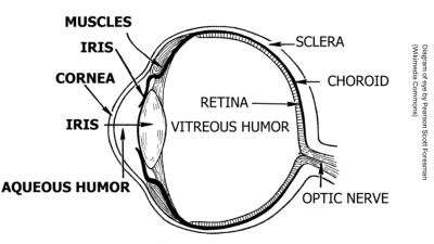
The vitreous is a transparent gel-like structure that lies behind the iris (the coloured part of your eye) and lens. It sits in front of the retina. The retina is the light sensing tissue at the back of the eye.
Choroidal biopsy
We remove a small part of the choroid (and retina).
The choroid is the tissue that forms the middle layer of the wall of the eye. It lies between the retina and the sclera (white outer layer of the eye). The choroid has a rich supply of blood vessels which bring oxygen and nutrients to the eye.
Why we recommend a biopsy for you
The biopsy helps us to plan the best management for your condition.
After we perform the biopsy, we send the sample of removed tissue to the laboratory where it is examined under a microscope to look for the presence of abnormal cells. The lab can also perform other tests on the sample.
During the biopsy
For both types of biopsy, we perform a vitrectomy. A vitrectomy is an operation to remove vitreous gel in the eye. This allows us to collect a sample of the vitreous and gain access to the retina and the choroid.
What happens to the vitreous gel
We do not replace your vitreous gel. We substitute your vitreous with one of the following substances after the vitrectomy:
- air (naturally absorbed within 1 to 7 days)
- a gas bubble (naturally absorbed within 2 to 10 weeks)
- silicone oil (this does not absorb and we may need to remove it at a later date)
Once the substance (air or gas) absorbs, the vitreous cavity gradually fills with aqueous humour (a clear fluid which is constantly produced by the eye).
Sedation during the operation
We usually perform the biopsy while you are sedated with local anaesthetic. This means you are awake during the operation, but your eye is numbed.
We give you medicine in your veins to help you feel more relaxed. Occasionally we perform the biopsy while you are under general anaesthetic (asleep).
Risks and side effects
Risks
The main risks of the biopsy include the following:
- serious eye infection (endophthalmitis). This affects 1 in 1000 patients. We can treat this with oral (by mouth) and local antibiotics (usually eye drops; sometimes we add ointments).
- retinal tears or detachment. We can treat these with laser or surgery, as required.
- increase in eye pressure. In most cases we can manage this with eyedrops or tablets. In rare cases we may offer surgery.
- inflammation or bleeding in the eye. We can treat this with eyedrops or tablets.
- cataract (cloudiness of the natural lens of the eye). This is a risk unless you have already had cataract surgery.
Side effects
Common side effects include:
- Red eye due to mild bruising on the surface of the eye. This clears in 1 to 2 weeks.
- Sore and gritty eye due to disturbance to the eye surface. This clears in 1 to 2 days.
Cataracts
Most patients who have a vitrectomy develop a cataract, affecting their vision, an earlier age than they would have otherwise. This may mean they need a cataract operation.
If a cataract is already present, cataract surgery may be performed at the same time as the vitrectomy.
After the biopsy
You can expect the following after the biopsy.
- Your vision will be blurry for a few weeks following surgery.
- If you have a gas or oil bubble in your eye, your vision will be blurry until the gas bubble naturally reabsorbs, or we remove the oil.
- You may notice a ‘curved edge’ of the air or gas bubble in your line of vision as the bubble gradually reabsorbs.
- We will give you eye drops to use following the surgery, to help to reduce inflammation and prevent infection.
- Sometimes we prescribe additional drops or tablets to keep your eye pressure low.
- Your eye is likely to be red for up to 4 weeks.
Everyday activities after the biopsy
You must avoid contact sports and swimming for 1 month after your biopsy.
If you require a general anaesthetic for another operation during this time, you must let the anaesthetist know that you have a gas bubble in the eye.
Flying
You must not fly whilst you have air or gas in your eye, as it may expand due to aircraft atmospheric pressure.
This can lead to a dangerous rise in your eye pressure.
Time off work
You will need to take approximately 2 weeks off work (depending on your job) to recover after your vitrectomy.
Driving
This depends on the vision in your other eye. Your doctor will advise you.
Follow up
It can take up to 2 weeks for lab results to come back. When they are back, we will invite you for a post-operation review, and share the results with you.
Contact information
Kingston Hospital Royal Eye Unit, Monday to Friday 9am to 5pm
Moorfields at St George's Hospital (Duke Elder Ward), Monday to Friday 4.30pm to 8.30am, 24 hours on weekends. Referral via an emergency GP or booked appointment required.